Ventilator
Settings
Ventilator settings are ordered by
the physician and are individualized for each patient. Ventilators are designed
to monitor many components of the patient’s respiratory status. Various alarms
and parameters can be set to warn healthcare providers that the patient is
having difficulty with the settings.
Respiratory
Rate (RR)
The respiratory rate is the number of
breaths the ventilator delivers to the patient each minute. The rate chosen
depends on the tidal volume, the type of pulmonary pathology, and the patient’s
target PaCO2. The respiratory rate parameters are set above and
below this number and the alarm will then sound if the patient’s actual rate is
outside of the desired range.
(The following are guidelines.) For
patients with obstructive lung disease, the rate should be set at 6-8
breaths/minute to avoid the development of auto-PEEP and hyperventilation, or
“blowing off CO2”. Patients with restrictive lung disease usually
tolerate a range of 12-20 breaths/minute. Patients with normal pulmonary
mechanics can tolerate a rate of 8-12 breaths/minute. The patient should be
monitored on the initial rate setting and adjustments made as necessary.
Tidal
Volume (VT)
The tidal volume is the volume of gas
the ventilator delivers to the patient with each breath. The tidal volume
parameters are set above and below the desired number, and the alarm will sound
if the patient’s actual tidal volume is outside of the desired range. This is
especially helpful if the patient is breathing spontaneously between
ventilator-delivered breaths, since the patient’s own tidal volume can be
compared with the tidal volume delivered by the ventilator.
The usual setting is 5-15 cc/kg,
based on compliance, resistance, and type of pathology. Patients with normal
lungs can tolerate a tidal volume of 12-15 cc/kg, whereas patients with
restrictive lung disease may need a tidal volume of 5-8 cc/kg.
Fractional
Inspired Oxygen (FiO2)
The fractional inspired oxygen is the
amount of oxygen delivered to the patient. It can range from 21% (room air) to
100%. It’s recommended that the FiO2 be set at 1.0 (100%) upon the
initiation of mechanical ventilation to allow the patient to get used to the
ventilator without experiencing hypoxia. However, 100% oxygen should not be
used continuously for long periods of time because of the risk of oxygen
toxicity. Oxygen toxicity causes structural changes at the alveolar-capillary
membrane, pulmonary edema, atelectasis, and decreased PaO2. Once the
patient is stabilized, the FiO2 can be weaned down based on pulse
oximetry and arterial blood gas values. The FiO2 should only be as
high as is necessary to keep the PaO2 in the desired range.
Most ventilators have a temporary
100% oxygen setting that delivers 100% oxygen for only a few breaths. This
should always be used prior to and after suctioning; during bronchoscopy, chest
physiotherapy, or other stressful procedures; and during patient transport.
Inspiratory:
Expiratory (I: E) Ratio
The I: E ratio is usually set at 1:2
or 1:1.5 to approximate the normal physiology of inspiration and expiration.
Occasionally, a longer inspiratory than expiratory time is desired to allow
more time to oxygenate the patient’s lungs. This is called inverse ratio
ventilation, and will be discussed later.
Pressure
Limit
The pressure limit regulates the
amount of pressure the volume-cycled ventilator can generate to deliver the
preset tidal volume. Because high pressures can cause lung injury, it’s
recommended that the plateau pressure not exceed 35 cm H2O. If this
limit is reached, the ventilator stops delivering the breath and alarms. This
may be an indication that the patient’s airway is obstructed with mucus, in
which case, the high pressure is usually resolved with suctioning. It can also
be caused by the patient coughing, biting on the ETT, breathing against the
ventilator, or by a kink in the ventilator tubing.
Flow
rate
The flow rate is the speed with which
the tidal volume is delivered. The usual setting is 40-100 liters per minute.
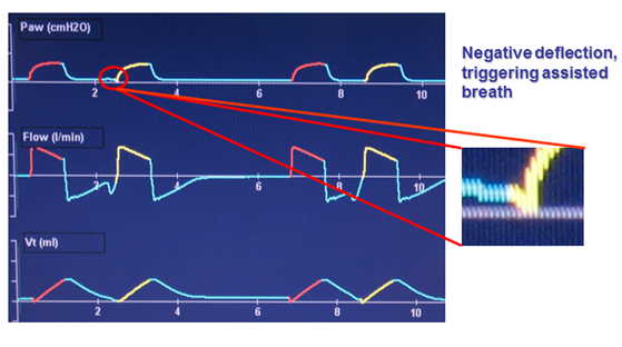
Sensitivity/Trigger
The sensitivity determines the amount
of effort required by the patient to initiate inspiration. It can be set to be
triggered by pressure or flow. Flow triggering is a better setting for patients
who can breathe spontaneously because it reduces the work of breathing.
Sigh
The ventilator can be
programmed to deliver an occasional sigh with a larger tidal volume. The use of
frequent sighs was popular during the 1970s because it was thought that it
prevented collapse of the alveoli (atelectasis), which can result from the
patient constantly inspiring the same volume of gas. However, recently there
has been concern that the increased pressure produced in the alveoli may
heighten the risk of the alveoli rupturing and causing pneumothorax.
Ventilator
Settings summary that nurses deal with the most
|
S.No.
|
SETTING
|
FUNCTION
|
USUAL
PARAMETERS
|
-
|
Respiratory
Rate (RR)
|
Number of breaths delivered by the ventilator per minute
|
Usually 4-20 breaths per minute
|
-
|
Tidal
Volume (VT)
|
Volume of gas delivered during each ventilator breath
|
Usually 5-15 cc/kg
|
-
|
Fractional
Inspired Oxygen (FiO2)
|
Amount of oxygen delivered by ventilator to patient
|
21% to 100%; usually set to keep
PaO2 > 60 mmHg or SaO2 > 90%
|
-
|
Inspiratory:Expiratory
(I:E) Ratio
|
Length of inspiration compared to length of expiration
|
Usually 1:2 or 1:1.5 unless inverse ratio ventilation is
required
|
-
|
Pressure
Limit
|
Maximum amount of pressure the ventilator can use to
deliver breath
|
10-20 cm H2O above peak inspiratory pressure; maximum is 35
cm H2O
|
Modes of
mechanical ventilation
Modes of mechanical ventilation are
described by the relationships between the various types of breaths and by the
variables that can occur during the inspiratory phase of ventilation. Each mode
of ventilation is distinguished by how it initiates a breath (trigger), how it
sustains a breath (limit), and how it terminates a breath (cycle); these are
referred to as phase variables.
The best mode of mechanical
ventilation is the one that provides maximum therapeutic benefit with the
fewest side effects. Mode selection and individual ventilator settings are
geared towards the patient’s diagnosis and history as well as integrated data
from laboratory, radiology and physical examination.
Basic modes of ventilation
1.
Continuous
Mandatory Ventilation (CMV)
2.
Assist
Control (A/C) Ventilation
3.
Intermittent
Mandatory Ventilation (IMV)
4.
Positive
End Expiratory Pressure (PEEP)
5.
Continuous
Positive Airway Pressure (CPAP)
Continuous Mandatory
Ventilation (CMV)
·
CMV
completely controls the patient’s ventilation. The ventilator provides a
mechanical breath on a preset timing. Patient respiratory efforts are ignored.
·
This
is generally uncomfortable for children and adults who are conscious and is
usually only used in an unconscious patient.
·
In
this mode the ventilator delivers a mechanical breath with pre-set volumes at a
pre-set rate and a pre-set flow rate.
·
The
patient CANNOT generate spontaneous breaths, volumes, or flow rates in this
mode.
Fig: Diagram shows display of volume,
flow and pressure waveforms as seen in the CMV mode. The shaded areas marked
with “E” represent the expiratory phase.
Disadvantage
The major disadvantage of
CMV is that it is not synchronized with the efforts of the patient. When the
patient is “out of sync” with the ventilator, he attempts to exhale as the
ventilator is in the inspiratory phase. As a result, airway pressure builds to
abnormally high levels and the remainder of the inspiratory volume is not
delivered. This “bucking” causes a high-pressure alarm. Signs and symptoms of
ventilator dys-synchrony include:
•
Agitation
•
Diaphoresis
•
Tachycardia
•
Tachypnea
•
Paradoxical
thoraco-abdominal breathing pattern
•
Increased
PIP (peak inspiratory pressure)
Assist Control (A/C)
Ventilation
·
The
A/C mode is similar to CMV, but it allows the patient to trigger an assisted
breath at any time.
·
A/C
delivers the pre-set volumes at a pre-set rate and a pre-set flow rate in
response to the patient’s own inspiratory effort, but will initiate the breath
if the patient does not do so within the set amount of time.
·
The
patient CANNOT generate spontaneous volumes, or flow rates in this mode. All
delivered breaths, whether mandatory or patient-triggered, will be delivered by
the ventilator according to the set parameters. i.e. All breaths in the
assist-control mode receive the same FiO2 and tidal volume.
·
Hyperventilation
and respiratory alkalosis may result from occurrences that increase the
patient’s spontaneous rate such as anxiety or neurological factors. A high
sensitivity setting that causes the machine to cycle too frequently can also
cause this problem. An increased risk of air trapping with high respiratory
rates may also potentially occur with the A/C ventilation.
·
The
A/C rate is the minimum number of full ventilator breaths the patient will
receive. The actual respiratory rate is equal to the A/C rate plus any
patient-triggered breaths per minute.
·
This
mode is used for patients who can initiate a breath but who have weakened
respiratory muscles.
Intermittent Mandatory
Ventilation (IMV) & Synchronous IMV (SIMV)
IMV
·
IMV is
the most commonly used modes of ventilation.
·
In
this mode the ventilator delivers a preset rate, tidal volume (or inspiratory
pressure) and FiO2.
·
The
patient may also draw spontaneous breaths in-between mandatory breaths. Unlike
A/C, breaths that the patient takes spontaneously do not trigger or cycle the
ventilator.
·
Patient-initiated
breaths are completely spontaneous, neither assisted nor supported by the
ventilator.
SIMV
·
SIMV
was developed as a result of the problem of high respiratory rates associated
with A/C.
·
SIMV
delivers the preset volume or pressure and rate while allowing the patient to
breathe spontaneously in between ventilator breaths. Each ventilator breath is
delivered in synchrony with the patient’s breaths, yet the patient is allowed
to completely control the spontaneous breaths.
·
SIMV allows
the patient to generate spontaneous breaths, volumes, and flow rates between
the set breaths.
·
SIMV
is used as a primary mode of ventilation, as well as a weaning mode. During
weaning, the preset rate is gradually reduced, allowing the patient to slowly
regain breathing on his or her own.
Advantages
·
Maintains
respiratory muscle strength by avoiding muscle atrophy
·
Decreases
mean airway pressure
·
Facilitates
ventilator discontinuation – “weaning”
·
Decreased
chance of hyperventilation,
·
Decreased
atrophy of accessory muscles, and
·
Improved
distribution of gas throughout the lungs by the action of the diaphragm.
Disadvantages
·
This
mode may increase the work of breathing and respiratory muscle fatigue.
·
In IMV
mode the mechanical rate and spontaneous rate may asynchronous causing
“stacking” and that may cause barotrauma or volutrauma
Spontaneous Modes OR
Customized Adjuncts to Ventilator Modes
PEEP (Positive End
Expiratory Pressure)
·
According
to its purest definition, the term PEEP is defined as positive pressure at the
end of exhalation during either spontaneous breathing or mechanical
ventilation. However, use of the term commonly implies that the patient is
also receiving mandatory breaths from a ventilator.
·
One
method of improving the patient’s oxygenation without increasing the FiO2
is the use of PEEP. Basically, PEEP does not allow airway pressure to return to
zero at the end of expiration.
·
PEEP
is not a mode of ventilation in itself. It is an adjunctive therapy added to
other modes. It is intended to improve oxygenation, not to provide ventilation,
which is the movement of air into the lungs followed by exhalation
·
PEEP
is added to increase functional residual capacity (FRC) and allow for a
decrease in the FiO2. PEEP helps to prevent small airway and
alveolar collapse, improves alveolar ventilation and may decrease the work of
breathing (at low levels). PEEP facilitates oxygen diffusion at lower FiO2
levels, which is safer for the patient.
·
PEEP
of 5cm H2O pressure is referred to as “physiologic” PEEP because it
is equivalent to the effect of the closed glottis. Therapeutic PEEP usually
ranges from 10-30cm H2O in adults.
·
PEEP
is an effective therapy for disease processes involving atelectasis; it is a
cornerstone of therapy for ARDS.
Disadvantage
·
Decreased
cardiac output with or without hypotension occurs because PEEP increases
intra-thoracic pressure, which in turn decreases the venous return to the heart
(preload).
·
Potential
volutrauma and barotrauma,
·
Increased
intracranial pressure and
·
Potential
loss of tidal volume
Continuous Positive
Airway Pressure (CPAP)
·
CPAP
is similar to PEEP except that it works only for patients who are breathing
spontaneously.
·
CPAP
is PEEP with no set rate on the ventilator. CPAP is primarily used as a mode of
non-invasive mechanical ventilation. It is occasionally used in the final
stages of ventilator weaning, but has minimal application for the mechanically
ventilated patient.
·
Patients
on CPAP do not receive positive pressure breaths from the ventilator. All
breaths are initiated and ended by the patient; tidal volumes and pressures are
variable from breath to breath.
·
CPAP
can also be administered using a mask and CPAP machine for patients who do not
require mechanical ventilation, but who need respiratory support; for example,
patients with sleep apnea.
·
CPAP
aids in promotion of oxygenation in the same way PEEP does. It has no influence
on ventilation.
Advantage
·
Ventilator
can monitor the patient’s breathing and activate an alarm if something
undesirable occurs
·
Helpful
for improving oxygenation in patients with refractory hypoxemia and a low FRC
·
CPAP
setting is adjusted to provide the best oxygenation with the lowest positive
pressure and the lowest FiO2

|
S.No.
|
MODE
|
FUNCTION
|
CLINICAL USE
|
|
1.
|
Control
Ventilation (CV)
|
Delivers
preset volume or pressure regardless of patient’s own inspiratory efforts
|
Usually
used for patients who are apneic
|
|
2.
|
Assist-Control
Ventilation (A/C)
|
Delivers
breath in response to patient effort and if patient fails to do so within
preset amount of time
|
Usually
used for spontaneously breathing patients with weakened respiratory muscles
|
|
3.
|
Synchronous
Intermittent Mandatory
Ventilation
(SIMV)
|
Ventilator
breaths are synchronized with patient’s respiratory effort
|
Usually
used to wean patients from mechanical ventilation
|
|
4.
|
Pressure
Support Ventilation (PSV)
|
Preset
pressure that augments the patient’s inspiratory effort and decreases the work
of breathing
|
Often
used with SIMV during weaning
|
|
5.
|
Positive
End Expiratory Pressure
(PEEP)
|
Positive
pressure applied at the end of expiration
|
Used
with CV, A/C, and SIMV to improve oxygenation by opening collapsed alveoli
|
|
6.
|
Constant
Positive Airway Pressure
(CPAP)
|
Similar
to PEEP but used only with spontaneously breathing patients
|
Maintains
constant positive pressure in airways so resistance is decreased
|
|
7.
|
Independent
Lung Ventilation (ILV)
|
Ventilates
each lung separately; requires two ventilators and sedation/paralysis
|
Used
for patients with unilateral lung disease or different disease process in
each lung
|
|
8.
|
High
Frequency Ventilation (HFV)
|
Delivers
small amounts of gas at a rapid rate (60-100 breaths/minute); requires
sedation/paralysis
|
Used
for hemodynamic instability, during short-term procedures, or if patient is
at risk for pneumothorax
|
|
9.
|
Positive
End Expiratory Pressure
(PEEP)
|
Positive
pressure applied at the end of expiration
|
Used
with CV, A/C, and SIMV to improve oxygenation by opening collapsed alveoli
|
Alarms
and Common Causes
As mentioned earlier, the ventilator
is designed to monitor many aspects of the patient’s respiratory status, and
there are many different alarms that can be set to warn healthcare providers
that the patient isn’t tolerating the mode or settings. The following are
common ventilator alarms and their most frequent causes.
|
High
Pressure Limit
|
Low
Pressure
|
High
Respiratory Rate
|
Low
Exhaled Volume
|
|
·
Secretions
in ETT/airway or condensation in tubing
·
Kink
in ventilator tubing
·
Patient
biting on ETT
·
Patient
coughing, gagging, or trying to talk
·
Increased
airway pressure from bronchospasm or pneumothorax
|
·
Vent
tubing not connected
·
Displaced
ETT or tracheostomy tube
|
·
Patient
anxiety or pain
·
Secretions
in ETT/airway
·
Hypoxia
·
Hypercapnia
|
·
Vent
tubing not connected
·
Leak
in cuff or inadequate cuff seal
·
Occurrence
of another alarm preventing full delivery of breath
|
BIBLIOGRAPHY
Books
1.
Brunner
LS, Suddarth DS, Smeltzer SCO. Brunner & Suddarth’s textbook of
medical-surgical nursing. Philadelphia: Lippincott Williams & Wilkins;
2008. Page No. 739-754
2.
Nettina
SM, Lippincott Williams & Wilkins. Lippincott manual of nursing practice.
Philadelphia: Wolters Kluwer Health : Lippincott Williams & Wilkins;
2010. Page No. 255-267
3.
Longo
DL, Harrison T. Harrison’s principles of internal medicine. New York, N.Y.,
[etc.]: McGraw-Hill Medical; 2012. Page No-
4.
Colledge
NR, Walker BR, Ralston S, Davidson S. Davidson’s principles and practice of
medicine. Edinburgh; New York: Churchill Livingstone/Elsevier; 2010. Page
No. 194-198
Web
page
1.
Adult
Invasive Mechanical Ventilation.pdf [Internet]. [cited 2012 May 28]. Available
from: http://www.mecriticalcare.net/downloads/mv/AdultInvasiveMechanicalVentilation.pdf
2.
Adult
Ventilation Management Online Nursing Continuing Education Course [Internet].
[cited 2012 May 28]. Available from: http://www.corexcel.com/courses/Vent_Web_Handout.pdf
3.
Critical
Care Nursing Theory - Mechanical ventilation [Internet]. [cited 2012 May 28].
Available from: http://www.philadelphia.edu.jo/academics/abatiha/uploads/Mechanical%20ventilation.pdf
4.
Patient
Education Series American Thoracic Society Mechanical Ventilation [Internet].
[cited 2012 May 28]. Available from: http://patients.thoracic.org/information-series/en/resources/mechanical-ventilation.pdf
5.
Core
Topics in Mechanical Ventilation [Internet]. [cited 2012 May 28]. Available
from: http://www.csen.com/vent.pdf
6.
Fundamentals
of Mechanical Ventilation [Internet]. [cited 2012 May 28]. Available from: http://www.ventworld.com/resources/pdf/vwchat.pdf
7.
Mechanical
Ventilation Critical Care Clinic [Internet]. [cited 2012 May 28]. Available
from: http://www.scribd.com/doc/25317501/Mechanical-Ventilation-Critical
8.
Mechanical
Ventilation for Nursing.ppt [Internet]. [cited 2012 May 28]. Available from: http://wwwappskc.lonestar.edu/programs/respcare/Missy%27s%20website/Mechanical%20Ventilation%20for%20Nursing.ppt
9.
Mechanical
ventilation Skills and techniques Update 2011 [Internet]. [cited 2012 May 28].
Available from: http://pact.esicm.org/media/Mechanical%20vent%201Feb2011%20final.pdf
10.
Mechanical
Ventilation.ppt [Internet]. [cited 2012 May 28]. Available from: http://home.cmcvellore.ac.in/upcoming/CME/ppt/Mechanical%20Ventilation.ppt
11.
Mechanical
Ventilation.ppt [Internet]. [cited 2012 May 28]. Available from: http://www.mcgill.ca/files/emergency/Mechanical_Ventilation.ppt
12.
Modes
of Ventilation ppt [Internet]. [cited 2012 May 28]. Available from: http://faculty.mdc.edu/pslocum/RET%202284%20Mod%203.0%20Modes%20of%20Ventilation.ppt
13.
Nursing
care of the mechanically ventilated patient: What does the evidence say? pdf
[Internet]. [cited 2012 May 28]. Available from:
http://eprints.qut.edu.au/33268/1/coyerdarticle%5B1%5D.pdf
14.
Nursing
Care Of The Ventilated Patient [Internet]. [cited 2012 May 28]. Available from:
http://intensivecare.hsnet.nsw.gov.au/five/doc/nurse_care_V_swahs.pdf
15.
Principle
of Mechenical Ventilation [Internet]. [cited 2012 May 28]. Available from: http://www.cmia.org/images/ventilation.pdf
16.
Chapter
82 - Mechanical Ventilation [Internet]. [cited 2012 May 28]. Available from: http://kemt.fei.tuke.sk/Predmety/KEMT537_LE/_materialy/09-Biomedical%20Engineering%20Handbook,%20The%20-Volumes%201%20%26%202/ch082.pdf
17.
respiratory-failure-mechanical-ventilation.pdf
[Internet]. [cited 2012 May 28]. Available from: http://www.thoracic.org/clinical/critical-care/clinical-education/respiratory-failure-mechanical-ventilation.pdf
18.
Advances
In Mechanical Ventilation [Internet]. [cited 2012 May 28]. Available from: http://www.newportnmi.com/FileDownloads/GENERAL-tobinmechvent.pdf
19.
Standard
of Practice: Care of the Mechanically Ventilated Patient [Internet]. [cited
2012 May 28]. Available from: http://www.cc.nih.gov/ccmd/cctrcs/pdf_docs/Ventilator%20Management/01-SOP-Carof%20the%20Mech.pdf
20.
Invasive
Mechanical Ventilation ppt [Internet]. [cited 2012 May 28]. Available from: http://home.cmcvellore.ac.in/upcoming/CME/ppt/Invasive%20Mechanical%20Ventilation.ppt
21.
Management
of the mechanically ventilated. Patient in the emergency department [Internet].
[cited 2012 May 28]. Available from: https://notendur.hi.is/thorsj/tasota/ED_MV.pdf
22.
Mechanical
Ventilation: A Review and Update for Clinicians [Internet]. [cited 2012 May
28]. Available from: http://seminmedpract.com/pdf/hp_dec99_vent.pdf
23.
How to
Withdraw Mechanical Ventilation [Internet]. [cited 2012 May 28]. Available
from: http://www.aacn.org/WD/CETests/Media/CI1841.pdf
24.
Mechanical
ventilation: Weaning and extubation [Internet]. [cited 2012 May 28]. Available
from: http://www.opus12.org/uploads/O12-SCI-V01-N02-P13.pdf
25.
Withdrawal
of Ventilatory Support from the Dying Adult Patient [Internet]. [cited 2012 May
28]. Available from: http://jso.imng.com/jso/journal/articles/0203283.pdf